|
Advice Regarding Diagnostic
Quality of Digital Transmissions
Digital images sent for diagnosis
must be of satisfactory quality such that they can be
interpreted clearly and correctly. Attempting to interpret
unsatisfactory digital images may be misleading and
inconclusive. In an effort to enhance your Second Opinion
experience, we provided some examples of images that may not
be readily suitable in order to avoid disappointment and delay
in a fitting response because resubmission of new digital
material is required.
 |
Insufficient Data – this image
does not include the entire tooth and related
structures. |
|
Poor angulation –
foreshortening
of the teeth results when images are taken at steep
angles, compressing the anatomical appearance. |
|
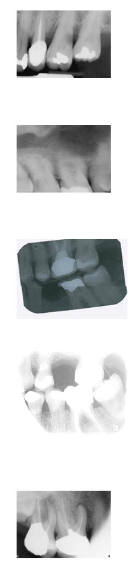
Bitewings – these are useful
for diagnosing caries or decay, but do not show the
complete anatomical structure of the teeth
|
|
Poor contrast and brightness –
diagnostic quality is compromised by a lack of
detail.
|
|
Cone cutting – part of the
image appears blank because of poor alignment of the
x-ray beam and the digital receiver. In some cases,
the area cone cut may be insignificant and the
radiograph can still be diagnostically useful.
|
| |
| |
We endeavor to with make your experience
with our service as pleasant and simple as possible by
illustrating some of these issues from the outset. We can only
help where you have helped us! |